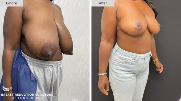
Some before photos speak for themselves before any history is given. This is one of those cases. The size, shape, and position of the breast tissue in the before photo tell a clear story about what this patient was carrying, and they explain exactly why surgery was needed.
Reading the Before Photo
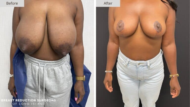
In the before photo, the breasts hang low and project far forward from the chest wall. The nipple-areola complex sits well below the breast fold, and the tissue has an elongated, stretched shape rather than a rounded one. This combination is a clear sign of significant volume paired with long-term stretching of the skin envelope.
The side view makes this even more obvious. From this angle, the breast tissue extends forward well past the chest, with very little support holding it in place. This kind of projection puts constant strain on the back, shoulders, and neck, since the weight is pulling away from the body instead of resting close to it.
Photos like this give us a lot of information before we even discuss symptoms or goals. The position of the nipple-areola complex alone tells us how much the support structures inside the breast have weakened over time. The deeper the fold between the breast and chest wall, the more it confirms how much the tissue has stretched and dropped.
What the Before Photo Tells Us About the Surgical Plan
When the before photo shows this level of volume and elongation, it shapes the entire surgical approach. A case like this needs more than a modest reduction. It requires removing a significant amount of tissue while also reshaping the breast mound and repositioning the nipple-areola complex much higher on the chest.
The elongated shape seen here also tells us the skin envelope has lost a lot of its elasticity. Skin that has been stretched this far for this long does not snap back on its own once tissue is removed. Our surgeon has to actively reshape and tighten the envelope during surgery, rather than relying on the skin to contract naturally afterward.
This is also where incision pattern becomes important. For breasts with this much volume and elongation, an anchor-shaped incision gives the access needed to remove tissue, reshape the lower pole, and reposition the nipple-areola complex all in the same procedure. Smaller incision patterns are not built to handle this level of correction.
Comparing the Before and After Side Profile
The side view comparison in this case is one of the clearest illustrations of how much the projection changed. In the before photo, the breast extends well beyond the chest wall. In the after photo, the breast sits close to the body, with a rounded, supported shape instead of a forward-hanging one.
This shift in projection is not just about removing volume. It comes from rebuilding the internal support of the breast so the remaining tissue sits higher and closer to the chest. Without this reshaping step, even a large reduction would leave the breast looking flat or under-supported.
The nipple-areola complex also moved significantly higher in the after photo, now sitting at a position consistent with a natural breast contour. This is a direct result of repositioning it during surgery rather than letting the skin alone determine where it ends up.
The Result
The after photos show a dramatic change in both size and shape. The breasts are smaller, sit higher on the chest, and have a rounded, proportional contour that was not visible in the before photos. The nipple-areola complex now sits in a natural position relative to the breast mound, and the forward projection seen before surgery has been replaced with a profile that sits close to the chest wall.
The symmetry between both sides has also improved, with both breasts matching closely in size, shape, and position. This kind of result reflects a surgical plan that addressed volume, shape, and support all at once, rather than focusing on just one part of the problem.
If Your Before Photo Would Look Similar
If you look at your own chest and recognize the same patterns described here, low nipple position, forward projection, or breasts that feel disproportionate to your frame, you may be a candidate for the same kind of correction. A before photo like this is often the clearest sign that a significant reduction is the right next step.
Every surgical plan is built around the individual anatomy shown in each patient’s photos. If your breasts have reached a point where the size and weight feel unmanageable, we encourage you to contact Breast Reduction Surgeons of Long Island for a consultation. Our team can evaluate your case and explain what a customized plan would look like for you.